Tennis Elbow
Print PdfPRP Injection Patient information leaflet
Tennis Elbow (Lateral Epicondylitis)
This condition is charactersied by pain and tenderness on the outside of the elbow. It occurs as a result of repetitive strain and trauma on the attachment of the extensor tendons of the wrist (these tendons bend the wrist back) and of the fingers, at the lower and outer portion of the arm (the outer 'bump of the elbow).
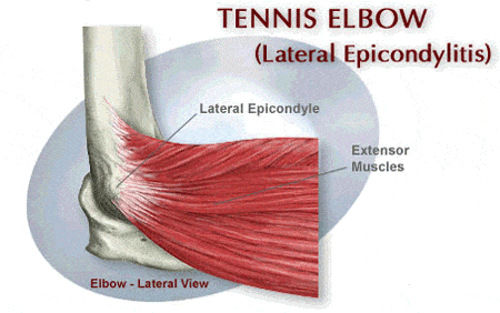
Fig 1: Shows the attachment of the extensor muscle at the outer part of the elbow which gets inflammed and painful.
Tennis elbow occurs as a result of repeated bending back of the wrist against resistance that leads to microtrauma and minor lesions and tears on the insertion of the extensor tendons depicted in the figure above.
Who is affected?
Although called Tennis Elbow at the end of the 19th century, this terms remains despite the fact that most of the people affected are not tennis players; instead other common causes are gardening, brick laying, excessive use of a screwdriver, hammering, computer typing and shaking hands.
What does tennis elbow feel like?
The main clinical symptom is pain centered on the lateral epicondyle (bony prominence on the outer aspect of the elbow) that radiates down into the forearm. The foremarm muscles may feel tight and sore. It is worsened by manouvres like lifting and gripping, especially so when the wrist is bent backwards. Tenderness just below the epicondyle and weakness of dorsal flexion of the wrist. Simple day to day actions like turning a door handle or picking up a bottle of milk can cause severe pain.
How is the diagnosis confirmed?
The diagnosis is mainly confirmed by clinical examination.
Normally plain X-Rays are not needed at the onset of the disease, but may be requested later on by the orthopaedic specialist to exclude other problems. An ultrasound scan may be performed if acute tendon tears are suspected or to visualize local sign of degeneration like calcific deposit that are associated with worse prognosis.
How is the condition treated?
Non-operative management
90% of the patients heal spontaneously within one year.
Conservative treatment is the treatment of choice for the first phase.
Avoiding repetitive wrist dorsiflexion (bending the wrist backwards) and modification of sport or offending activities are generally the most important prescriptions.
Pain killers and Non steroidal anti-inflammatory medications.
Local corticosteroid injections (up to 3 injections) are effective for short term pain control.
A Counterforce dynamic brace can be used; but there is poor patient compliance.
A physical therapy program (aimed to stretch and progressively strengthen the extensor muscles with pain free active and isometric exercise) has been shown to be effective in the long term.
PRP (Platelet Rich Plasma) injection
Platelet rich plasma (PRP) is blood plasma with concentrated platelets (the body’s repairmen for damaged tissue). The concentrated platelets found in PRP contain growth factors that are vital to initiate and accelerate tissue repair and regeneration. These bioactive proteins initiate connective tissue healing and repair, promote development of new blood vessels, and stimulate the healing process.
How does PRP therapy work?
Blood will be taken from you and then placed in a machine that spins at high speed to separate the different types of blood cells. The surgeon will extract the platelet rich part of the blood, mix it with local anaesthetic and inject this into the area of your injury. The entire process to prepare your blood takes about 15 minutes and increases the concentration of platelets and growth factors at the site of injury by up to 500% (you will have five times the normal number of platelets/growth factors). By having a PRP injection, we aim to stimulate your body’s ability to heal chronic conditions like tennis elbow.
What are the potential benefits of treatment?
The main benefit is that patients can see a significant improvement in symptoms. This treatment may eliminate the need for more aggressive treatments such as long term medication or surgery, as well as a remarkable return of function and a much shorter recovery time.
A major advantage of this treatment is that no foreign substance is used – we use the patient’s own growth factors from his or her own blood so there is no risk of any disease transmission.
What are the alternatives?
They include:
- Surgery
- Anti-inflammatory drug therapy
- Steroid injections
- Physiotherapy
What are the possible risks or complications of this procedure?
As with all surgery there is a risk of some complications. These are rare, but you should be aware of them before your operation. They include:
- Infection at the site of the injection.
- An increase in inflammation and pain at the site of the injection.
- Bleeding and/or bruising.
- No relief or worsening of symptoms.
- Skin discolouration.
- Allergic reaction to the local anaesthetic drug.
- Failure to achieve successful result.
- Injury to the nerves or blood vessels.
- Prolonged stiffness and or pain.
If you require further information about risks or complications, please discuss with the doctors in clinic or on admission.
How long will the procedure take?
The procedure usually takes around 30 minutes. Most of this time is separating the platelet-rich plasma from your blood sample.
This procedure is performed with the patient awake. The PRP is injected together with local anaesthetic drug.
Operative management
About 10% of cases are not responsive to conservative treatment. These patients could have symptoms more than 1 year of duration, more than 3 steroid injections in the past, constant pain without activity, local calcification or exostosis on XRay. In these cases surgery is a good option.
Patients satisfaction after surgery has been reported very high with more than 90% of good or excellent results.
3 different surgical options are available: open surgery, keyhole surgery, or per-cutaneous techniques.
Minimally invasive techniques like percutaneous or key hole techniques have been described as less harmful with quicker recovery and return to work activities. The key- hole technique also has the advantage to recognize and treat associated intra-joint abnormalities, if they are present.